| |
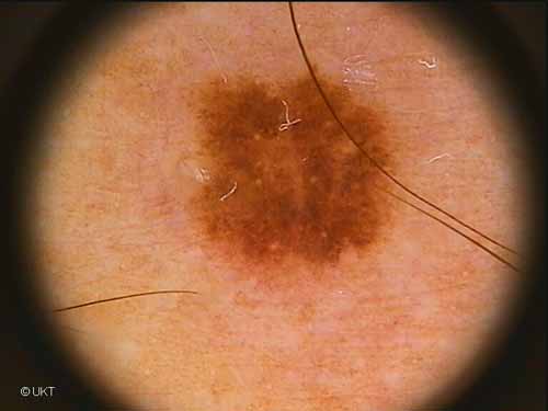
52-year-old male patient
Localisation: chest
Dermatoscopy: Symmetric lesion with graduated and irregular edge.
In the periphery relatively little pigmentation, towards the centre
uniform homogeneous pigmentation with a central depigmentation. Some
milia-like cysts.
Histology: Melanocytic compound nevus with dysplasia, lentiginous
hyperplasia, basal hyperpigmentation and regular junctional nests.
Auswahl Moleanalyzer
I
Orginal Moleanalyzer
II
|