| |
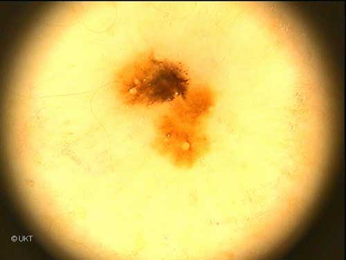
31-year-old
male patient
Localisation: shoulder
Dermatoscopy: Asymmetric lesion with irregular, graduated and
abrupt ending edge. In the lower region homogeneous brown pigmentation.
In the upper region brown to black pigmentation with globules and dots.
In the region of dark areas radial streaming and pseudopods. Some milia-like
cysts and follicular plugs.
Histology: Melanoma in situ with pronounced pagetoide spread
of melanocytes within the epidermis and hyperpigmentation. Melanin pigments
in the stratum corneum.
Auswahl Moleanalyzer
I
Orginal Moleanalyzer
II
|