| |
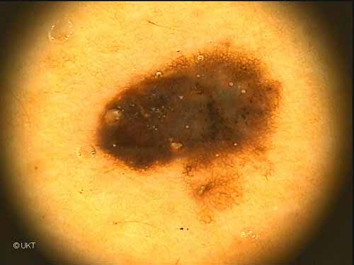
53-year-old male patient
Localisation: back
Dermatoscopy: Asymmetric lesion with both abruptly ending and
graduated edge. In the region of graduated edge regular network. Towards
the centre and on the left region of the lesion uniform homogeneous
dark brown to black pigmentation. In the centre faint grey veil. Some
dark globules and dots. Some milia-like cysts. One big cyst on the left
side of the lesion.
Histology: Superficial spreading melanoma, Breslow's tumour thickness
0.25 mm, Clark's level of invasion II.
Auswahl Moleanalyzer
I
Orginal Moleanalyzer
II
|