| |
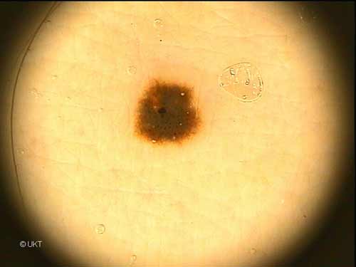
60-year-old male patient
Localisation: upper arm
Dermatoscopy: Symmetric lesion. Nearly abrupt ending edge. Regular
network with nearly homogeneous pigmentation in the centre. Multiple
black dots. Some milia-like cysts.
Histology: Lentiginous nevus with lentiginous hyperplasia and
basal to suprabasal hyperpigmentation. In the central area beginning
junctional nest formation is present. Scattered melanophages are seen
in the upper dermis.
Auswahl Moleanalyzer
I
Orginal Moleanalyzer
II
|