| |
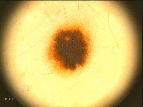
16-year-old
female patient
Localisation: back
Dermatoscopy: Nearly symmetrical lesion with graduated red-brown
edge. Regular network with homogeneous pigmentation in the centre. Some
black globules and dots. An area of light pigmentation above the centre.
Some milia-like cysts.
Histology: Spitz nevus with a junctional component and deeply
hyperpigmented nests with spindle-shaped cells. Many melanophages are
present in the upper dermis.
Auswahl Moleanalyzer
I
Orginal Moleanalyzer
II
|