| |
52-year-old female patient
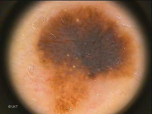
Localisation: shoulder
Dermatoscopy: Asymmetric lesion with graduated edges. Upper region
of uniform homogeneous pigmentation (colour blue-grey to brown, resembling
a blue-white veil). Some radial streaming-like areas and some milia-like
cysts. In the lower region nearly homogeneous brown pigmentation with
globules. The dermatoscopic differential diagnosis: superficial spreading
melanoma.
Histology: Seborrhoic keratosis with papillomatosis, horn cysts
and hyperplasia of melanocytes and hyperpigmentation. Subtype of melanoacanthoma.
Auswahl Moleanalyzer
I
Orginal Moleanalyzer
II
|